Johnson & Johnson Institute
Laparoscopic cholecystectomy
Relevant anatomy
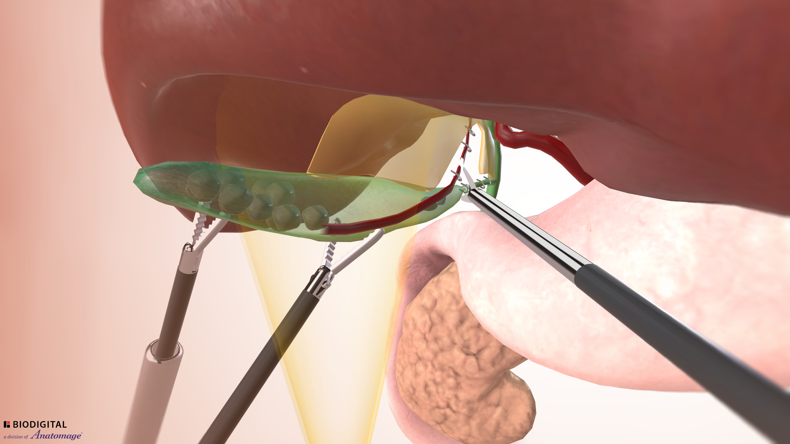
Click to interact with a BioDigital Human animated 3D anatomy, disease states, and procedure tour.
Key anatomical structures and landmarks:
- The triangle of calot (Hepatocystic Triangle) is anatomical space located near the porta hepatis. It is an area of focus during cholecystectomy.
- The triangle of calot is defined by three structures:
- Superior: inferior surface of the liver
- Lateral: cystic duct
- Medial: common hepatic duct
- The cystic artery normally resides within the triangle of calot.
References
George Crawford, MD prepared this procedure guide on behalf of Johnson & Johnson and its affiliates. The procedure guide reflects the opinions of the individual presenter, and the steps described may not encompass the complete steps of the procedure. Additionally, other surgeons may prefer different techniques, approaches, etc., as individual surgeon experience in his/her clinical practice, as well as patient needs, may dictate variation in procedure steps.
Before using any medical device, review all labeling, including without limitation; the Instructions For Use (IFU), and relevant package inserts with particular attention to the indications, contraindications, warnings and precautions, and steps for use of the device(s).
This presentation is not accredited for CE/CME.
George Crawford, MD is compensated by and presenting on behalf of Johnson & Johnson and its affiliates and must present information in accordance with applicable regulatory requirements.
US_ETH_WOUN_117866