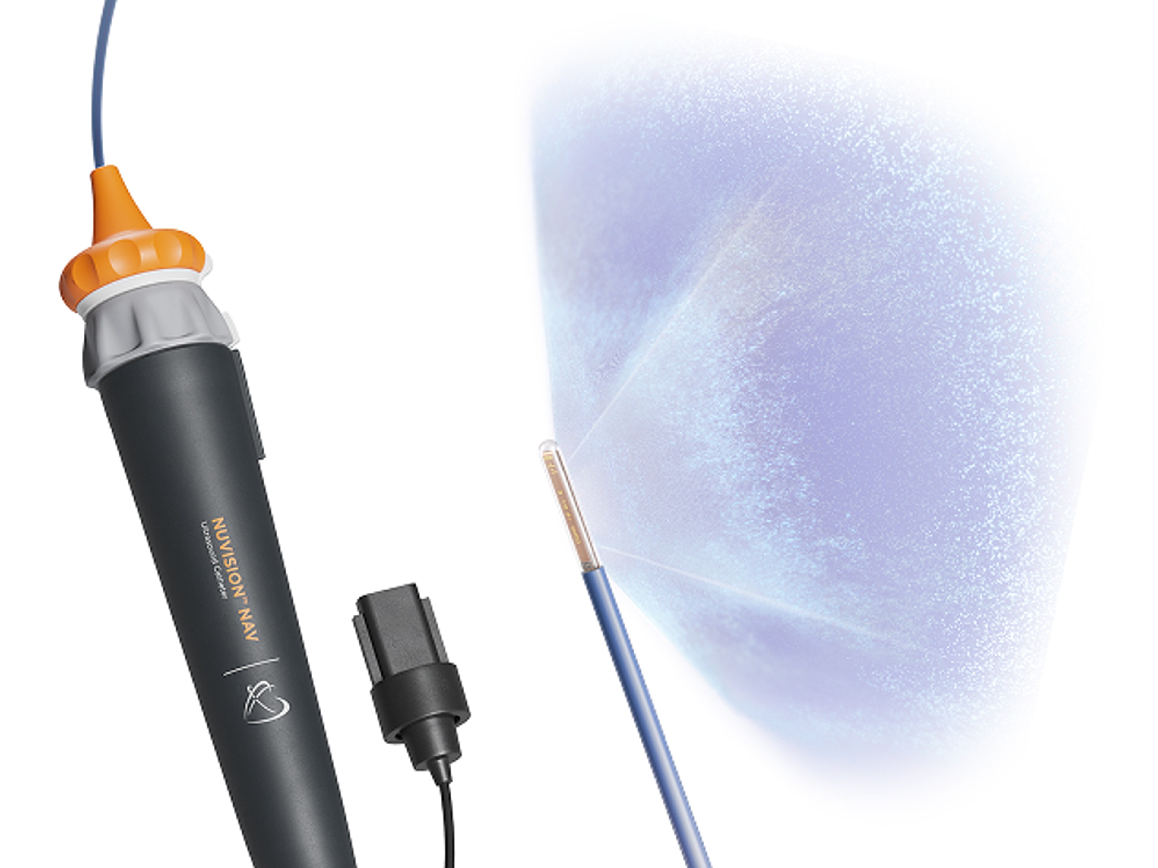
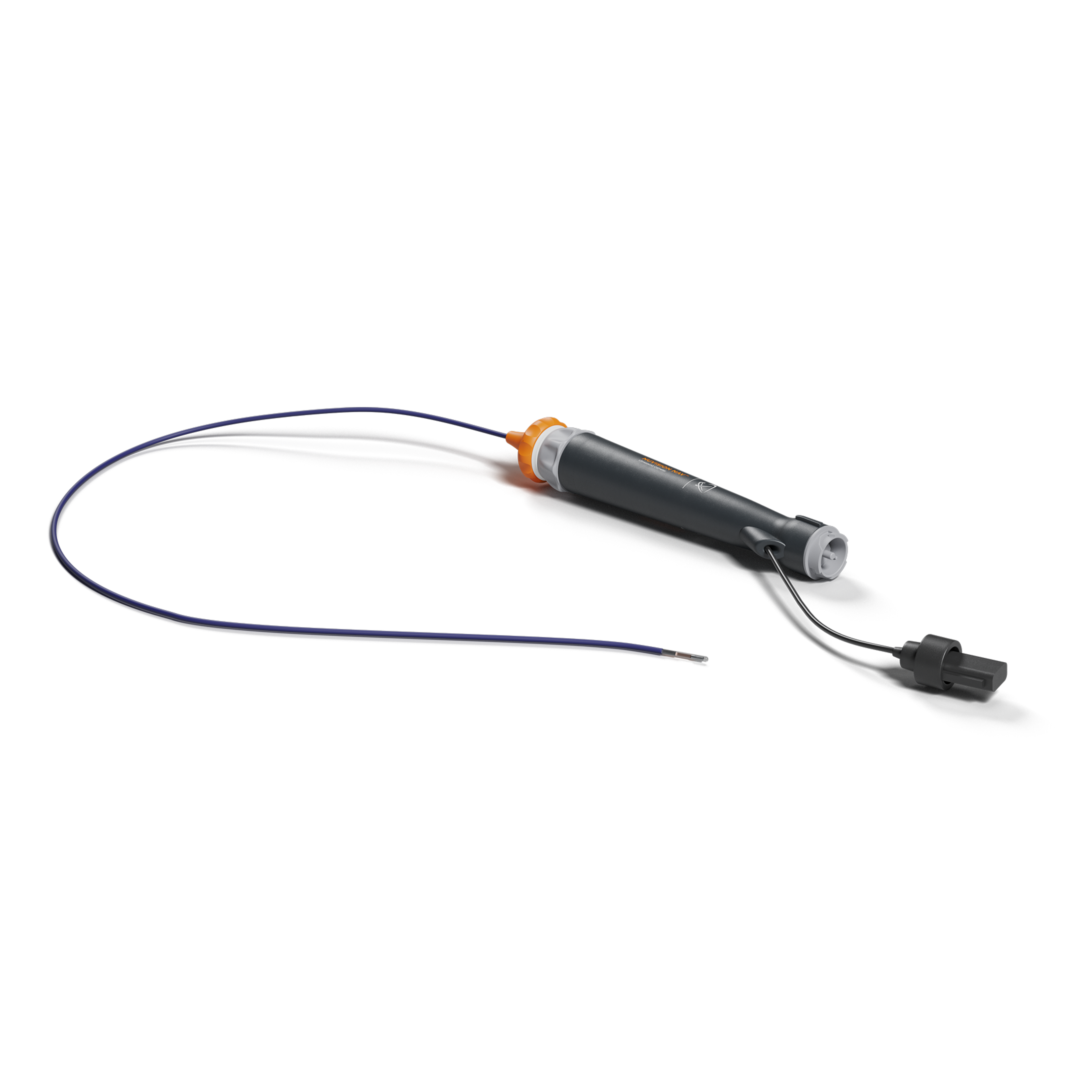
NUVISION™ NAV Ultrasound Catheter
NUVISION™ NAV Ultrasound Catheter is the most comprehensive 4D ICE guidance available for concomitant procedures.
About this device
Only NUVISION NAV™ seamlessly integrates 4D ICE ultrasound with CARTO™ 3 System, the world’s leading cardiac mapping system, to provide real-time high-definition tissue visualization.
By elevating integration to include 4D ICE, physicians can confidently and efficiently navigate procedures with a new level of visualization, only from Johnson & Johnson MedTech, your expert partner in ICE.**
Specialties
Cardiac Electrophysiology (EP)
Procedures
Cardiac Catheter Ablation, Intracardiac Echocardiography (ICE), Left Atrial Appendage Occlusion (LAAO)
Experience the best of both worlds
With CARTO™ 3 System integration, the single operator view and digital steering of the NUVISION™ NAV Catheter image through the remote user interface allows for a hands-free ICE catheter workflow.*
The versatility your concomitant EP procedures demand
NUVISION™ NAV Ultrasound Catheter enables you to do more in a single procedure, with the ability to guide ablation and structural heart procedures using the same 4D ICE catheter.
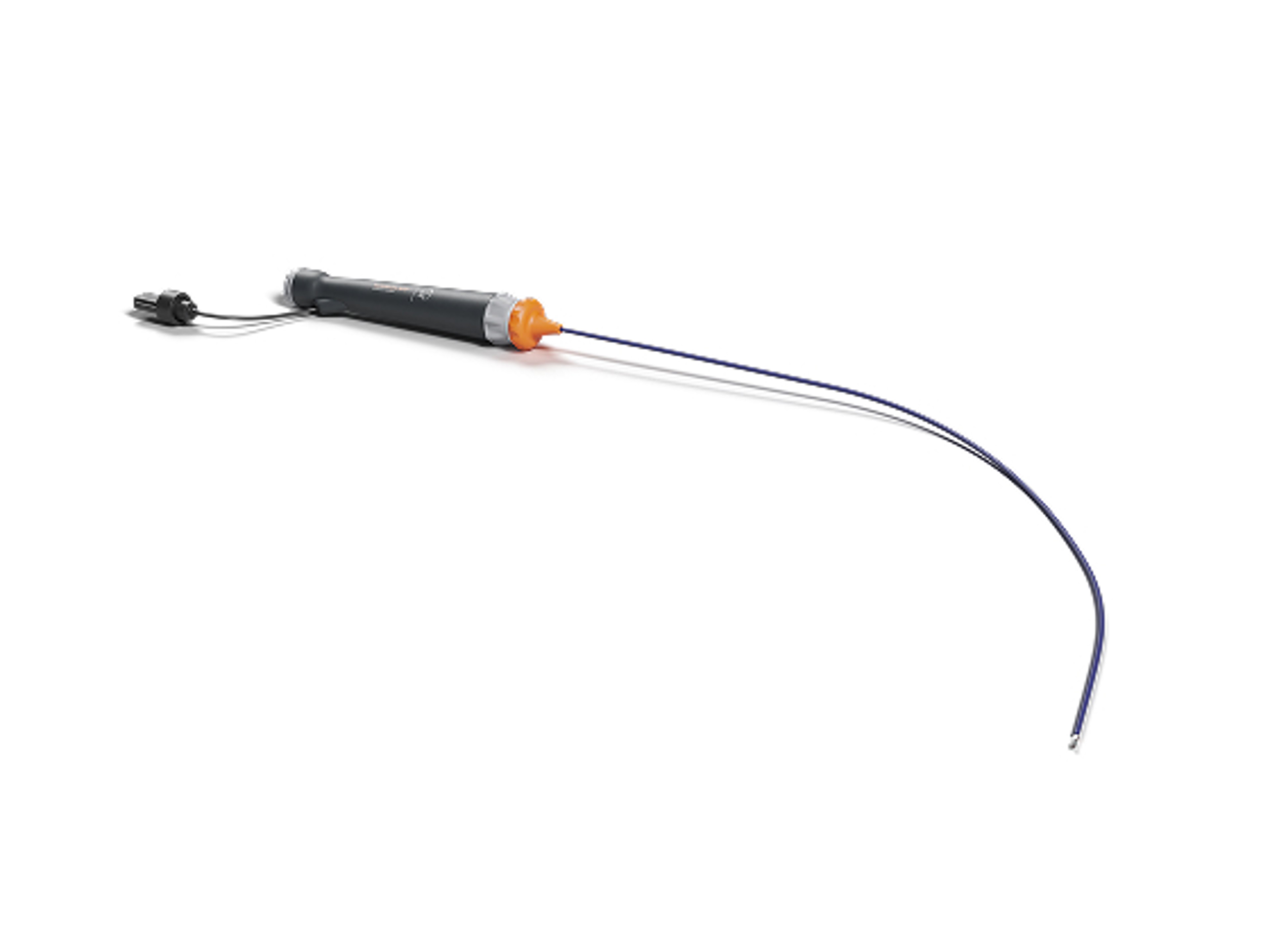
- 10.5Fr x 90cm torqueable and deflectable shaft
- 360° independent rotating tip for more flexibility in imaging acquisition
- Signature tip can be deflected up to +120° / -120°
96%
Success in device selection
NUVISION™ Ultrasound Catheter measurements accurately predicted the final device size that was implanted in 96.3% of cases.1,i
Registry study (n=274); NUVISION™ Ultrasound Catheter group 96.3% vs. 2D-ICE group 80.4% (p=0.005)
20%
Better prediction compared to 2D ICE
Enjoy high-definition, real-time guidance for both interventions, without the need to revert to TEE or perform catheter exchange.
28%
Shorter procedure time
Overall procedure time was significantly shorter when ICE was used to guide LAAO procedures, compared to TEE guidance***
Features & benefits
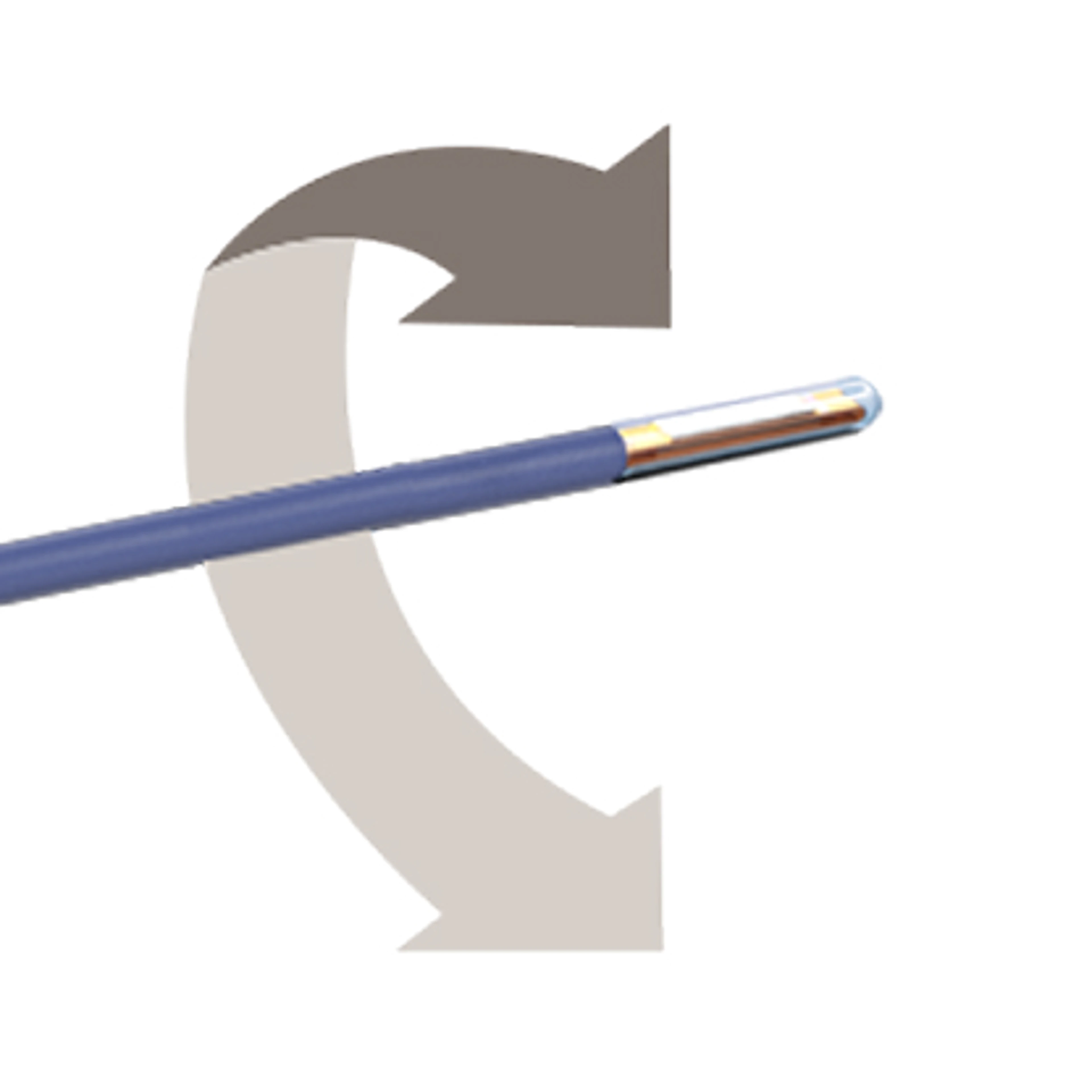
Advanced maneuverability at every turn
A 360° rotating tip and deflectable distal shaft enhances your abilities to map any anatomical structure with ease
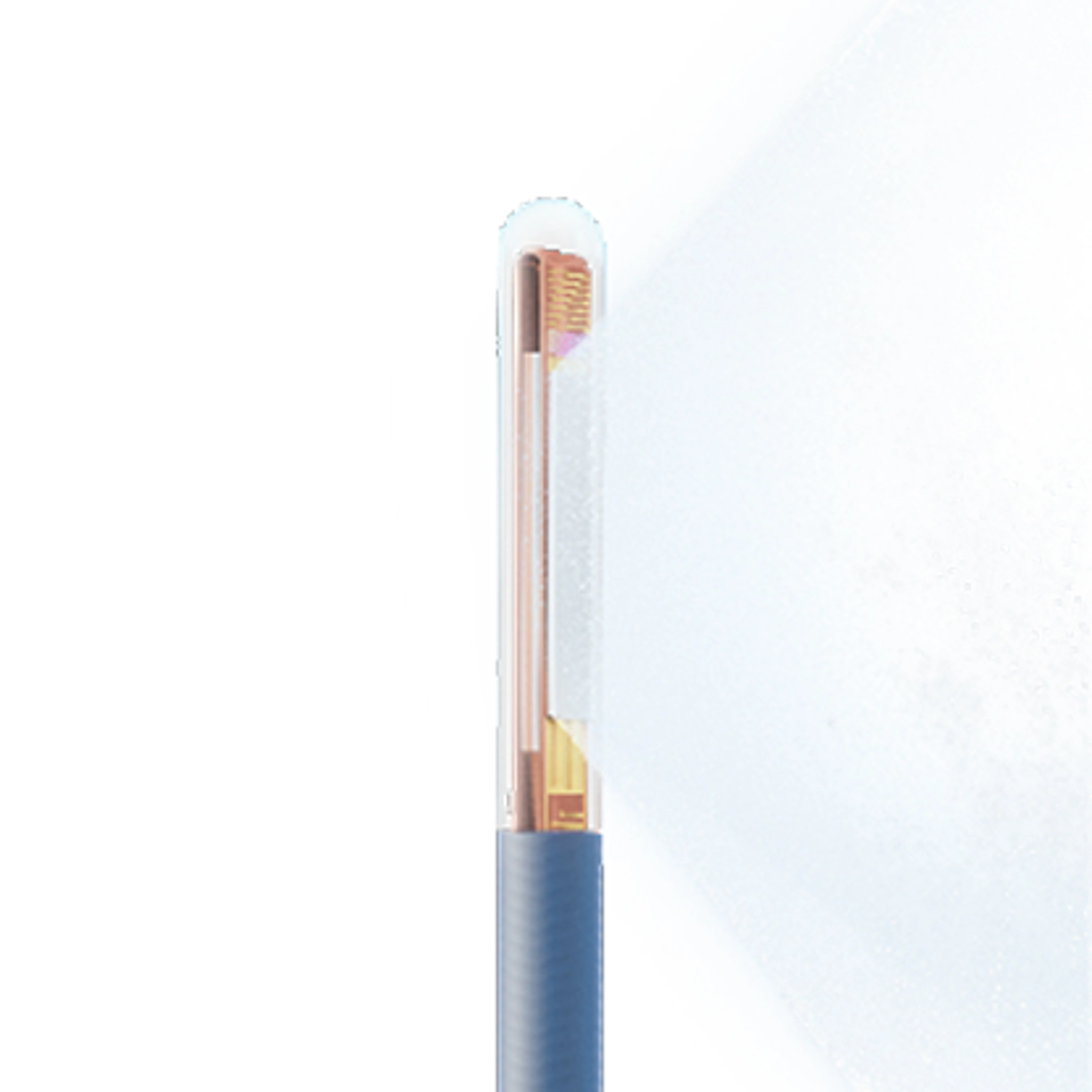
Multiplanar imaging
- 840 elements phased-array
- Wide 90° by 90° field of view with fast volume rates
- Azimuthal,elevation, coronal planes
- Ultrasound penetration depth up to 15cm
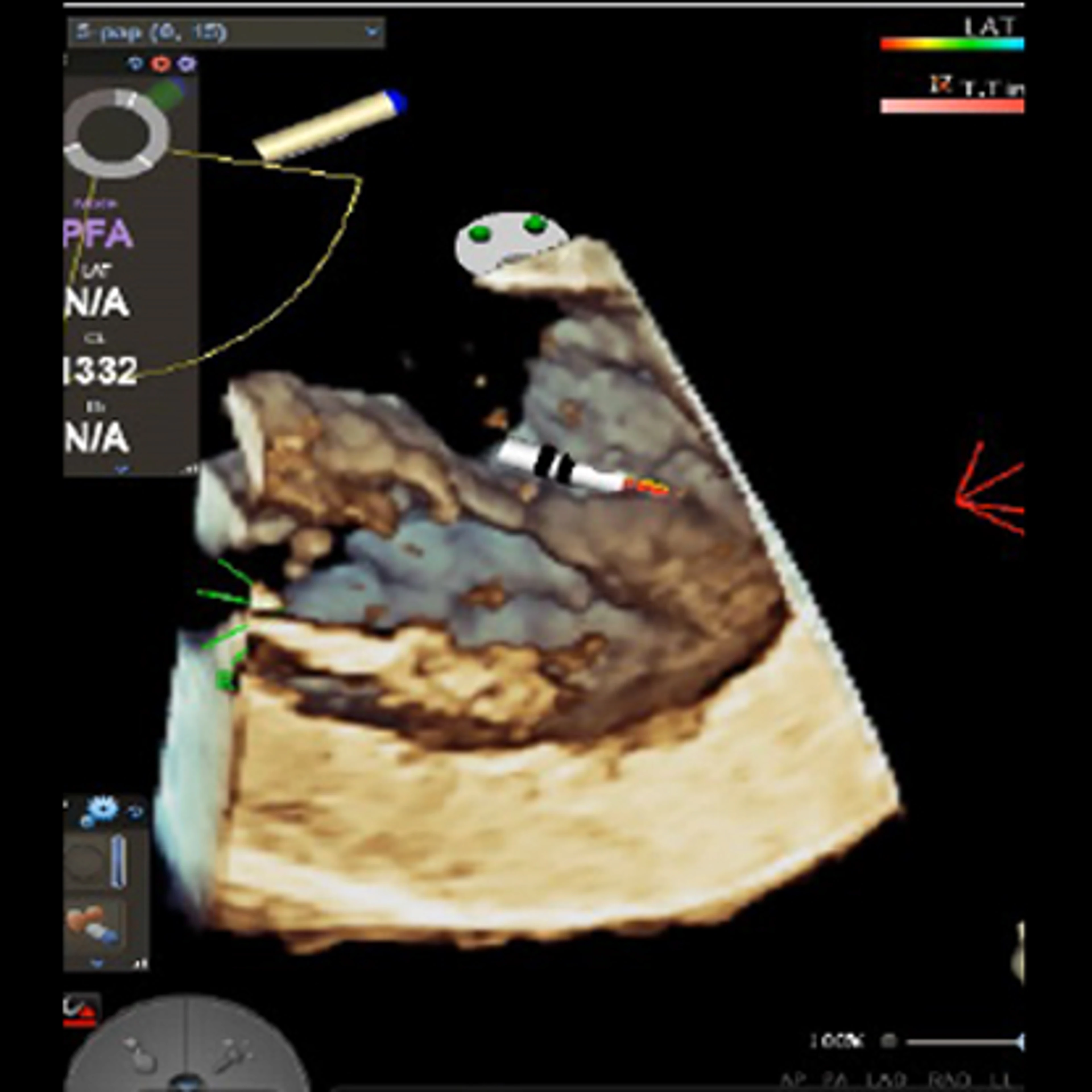
4D advanced mode
Enhances ultrasound data visualization and editing by allowing users to manipulate rotation, tilt, and depth of planes, customize image layouts, and utilize tools for cropping, contouring, and transparency control.
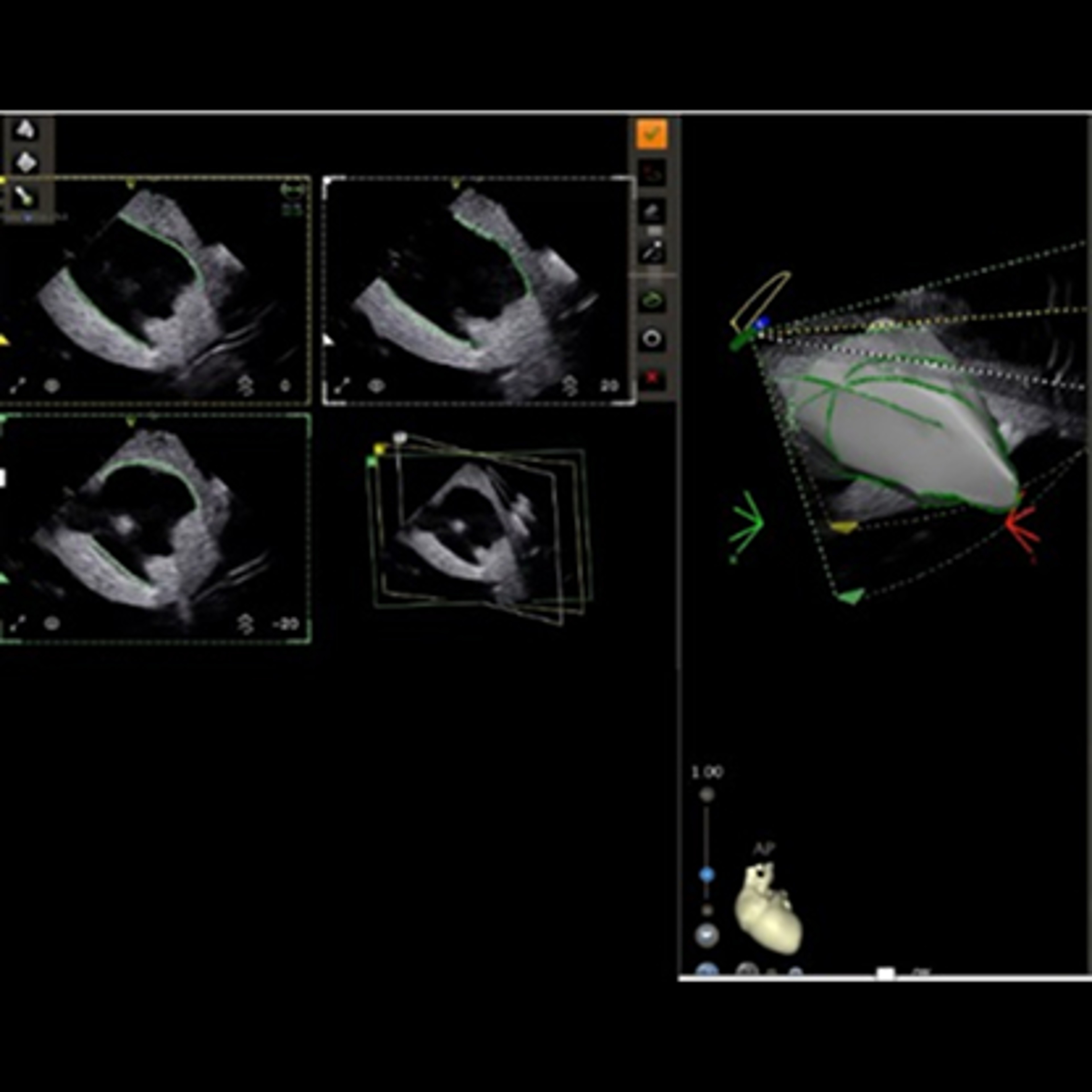
Multiplane CARTOSOUND™ Module
Includes Bi-plane and Tri-plane sub-modes, enhances efficiency in reviewing clips and creating contours. This innovative system supports a hands-free catheter workflow, streamlining your approach in all ventricular tachycardia cases.
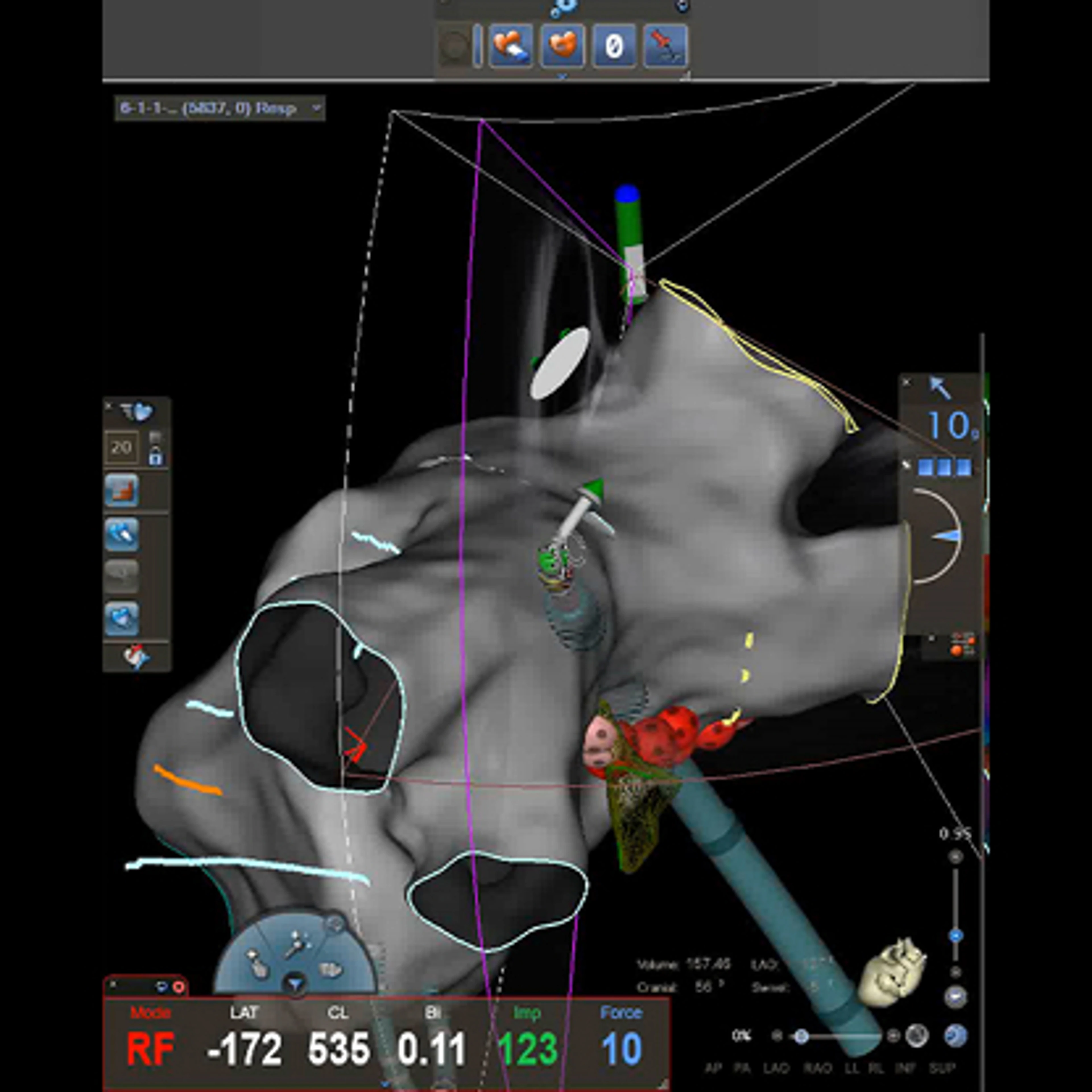
Ablation catheter tracking
Allows you to track the tip of your focal ablation catheters in any 2D imaging mode (Bi-Plane, Tri-Plane, V-Plane and 2D) to ensure you have tissue to catheter contact without the need to manipulate the NUVISION™ NAV Ultrasound Catheter.
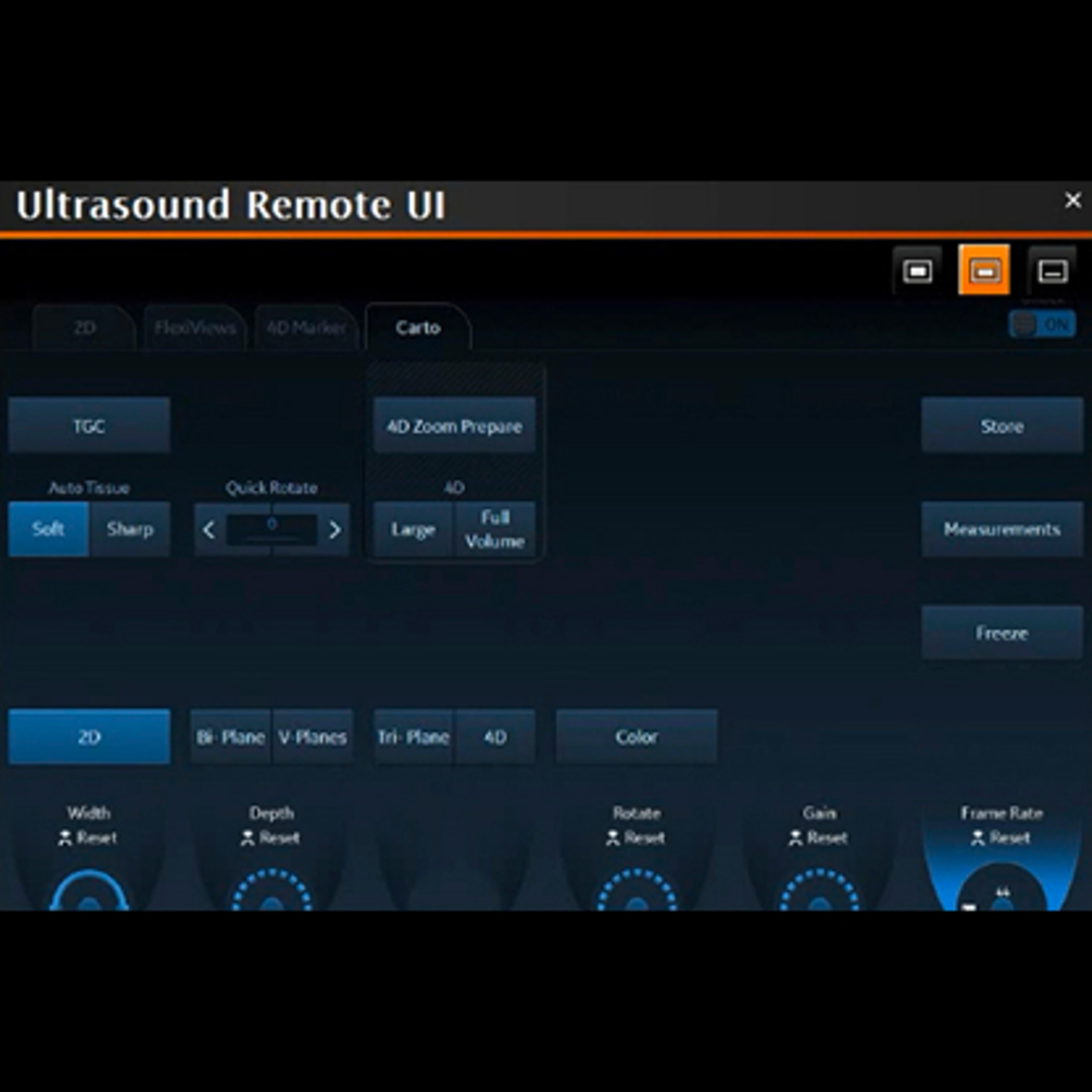
Remote User Interface
Enables control of the Vivid™ S70N Ultrasound System from the CARTO™ 3 System mapping system eliminating the need to control the ultrasound machine at the console, streamlining your procedural workflow.
Supporting documentation
Related products
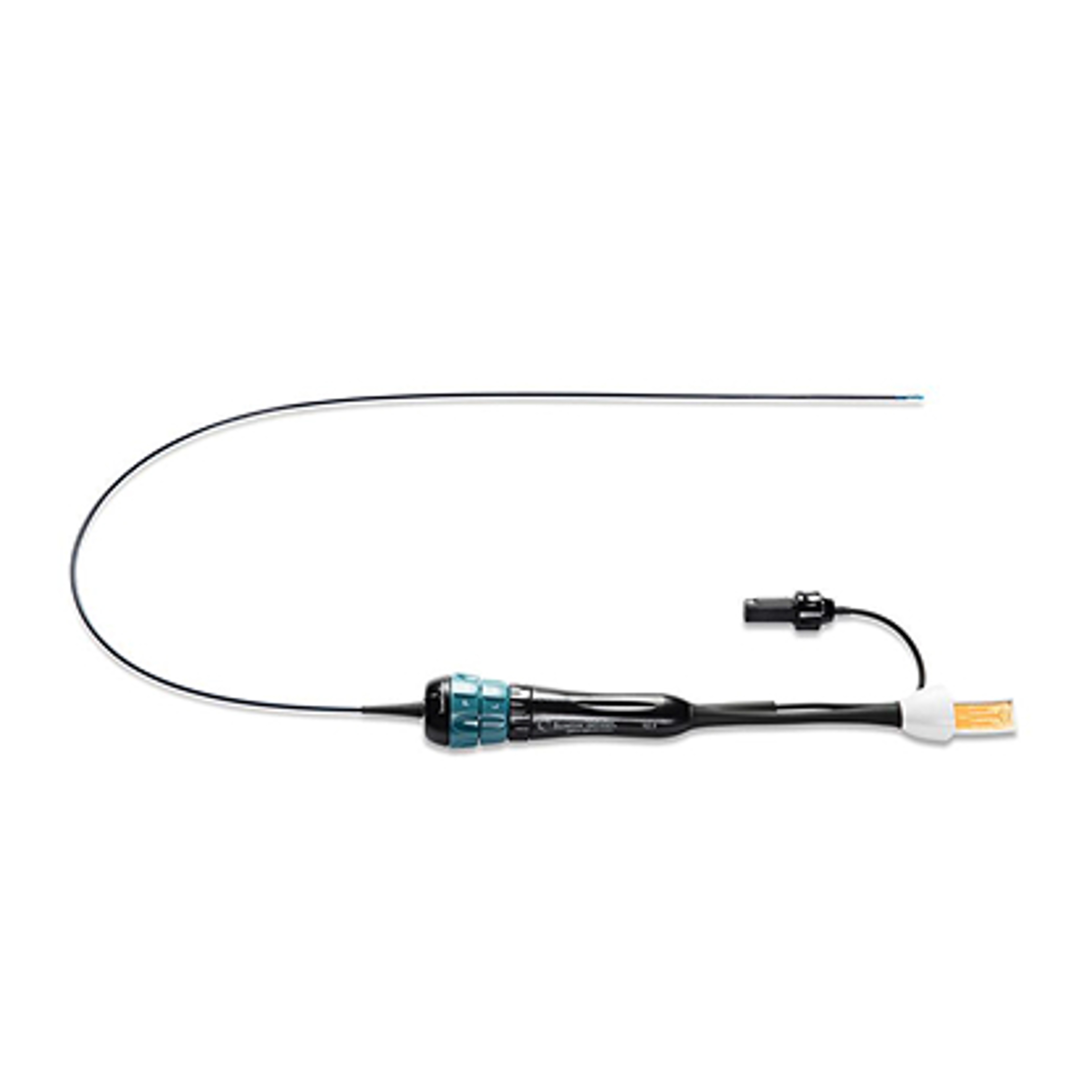
NUVISION™ Ultrasound Catheter
Break through to a new level of capability and confidence in structural heart procedures.
NUVISION™ Ultrasound Catheter is expertly designed to give you more. Better resolution for a comprehensive view of the heart -- and control, with its signature rotating tip -- for a simple, streamlined workflow.

SOUNDSTAR CRYSTAL™ Ultrasound Catheter
Integrated with the CARTOSOUND™ FAM Module, SOUNDSTAR CRYSTAL™ Ultrasound Catheter offers clear visualization and tissue definition with enhanced far field imaging, giving users added confidence in procedures and elevating the CARTO™ 3 System experience.
SOUNDSTAR™ Ultrasound Catheter
The SOUNDSTAR™ Ultrasound Catheter with the CARTOSOUND™ FAM Module delivers real-time intracardiac echocardiography imaging and navigation to enable workflow efficiencies, such as real-time monitoring of the ablation catheter tip to reduce radiation exposure and improve confidence.
References
* With the compatible CARTO™ 3 EP Navigation System.
** When compared to 2D Ultrasound
*** Retrospective study (n=144); ICE guided 58 min vs. TEE guided 80 min (p<0.001).
i. 2D ICE catheters used: 10 F SOUNDSTAR™ or AcuNav™ Ultrasound Catheter and 9 F ViewFlex Xtra ICE Catheter.
- Della Rocca DG, Magnocavallo M, Gianni C, et al. Three-dimensional intracardiac echocardiography for left atrial appendage sizing and percutaneous occlusion guidance. Europace. 2023;26(1):euae010
The third-party trademarks used herein are the trademarks of their respective owners.
Important information: Prior to use, refer to the instructions for use supplied with this device for indications, contraindications, side effects, warnings and precautions. Caution: US law restricts this device to sale by or on the order of a physician.
This site is published by Johnson & Johnson and its affiliates, which is solely responsible for its contents. It is intended for visitors from the United States. © Johnson & Johnson and its affiliates 2025 Last Updated on 09/15/2025.
US_ELP_ULTR_408139